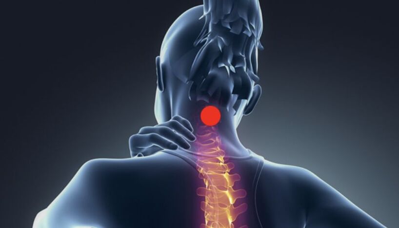
Causes of cervical osteochondrosis
- Overweight, sedentary lifestyle;
- Poor posture, scoliosis;
- Rheumatism;
- flatfoot;
- ventricular septal defect;
- Malnutrition.
General signs and symptoms
- Radicul syndrome - pain that spreads from the neck to the shoulder blades, forearms, and covers the front wall of the chest due to compression of the nerve endings;
- Arm muscles are weak and the neck is significantly swollen;
- You will hear a characteristic crunching sound when you move your head;
- Weakness, chronic fatigue, changes in blood pressure;
- Lack of coordination, frequent dizziness, nausea and vomiting during attacks;
- Deterioration of vision and hearing, noise, tinnitus;
- Numbness of limbs and tongue;
- Frequent migraines;
- Women aged 45-65 years old will experience pain, numbness and tingling in the upper limbs during sleep; the attacks can be repeated several times at night.
Classification of cervical osteochondrosis
- Grade 1 osteochondrosis - There are no particularly obvious symptoms in the initial stage. When turning and tilting the head, the patient will feel a rare slight pain, and the back muscles will soon tire.
- Secondary osteochondrosis - the vertebrae become unstable, nerves become pinched, unpleasant sensations in the neck become noticeable and radiate to the shoulders and arms. Other symptoms include increased fatigue, frequent headaches in the occipital area, and difficulty concentrating.
- Grade III Osteochondrosis - Pain becomes chronic and covers the upper back, arms, severe muscle weakness is observed, numbness of the limbs, intervertebral hernias develop, frequent attacks of dizziness occur.
- Grade 4 osteochondrosis - the intervertebral disc is completely destroyed and replaced by connective tissue, the pathological process covers several parts of the spine. Lack of coordination, more frequent attacks of dizziness, and ringing in the ears.
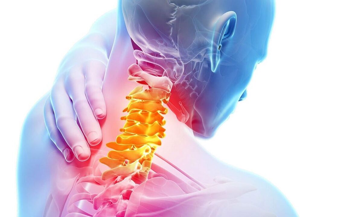
Which doctor should I contact?
diagnosis method
- X-ray– This method is effective only in the early stages of pathological development;
- MRI– The bone structure, the size and direction of the intervertebral hernia and the condition of the spinal cord are clearly visible on the screen;
- CT– This method is less effective than MRI because it does not provide accurate information about the presence and size of the hernia;
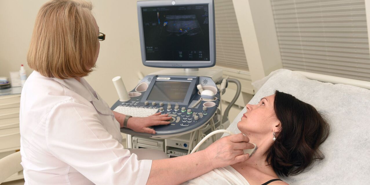
- Duplex scanning– Allows you to see blood flow disturbances;
- electroneurogram– Shows the presence of compression, inflammation and other nerve damage;
- Cerebral blood flow map– Treats blood supply problems to the brain.
Methods of treating cervical osteochondrosis
first aid
physiotherapy
- Maintain a standing position with your arms lowered freely along your body. Tilt your head forward, keep your chin against your chest as much as possible, and fix the position when you count to 3. Tilt your head back, look up with your chin, and count to three. Return to starting position.
- While standing, turn your head to the right, to the left, and hold the position for a few seconds at each extreme point. Repeat 3 times on each side.
- While standing, tilt your head to the right or left, try to rest your ears against your shoulders, and maintain this position for 30 seconds. Repeat 6 times in each direction.
- Stand with your hands on your waistband, chin parallel to the floor, and reach forward. Turn your head, rest your chin on your shoulders, rotate your torso slightly, and hold for half a minute. Repeat 6 times in each direction; slight pain may occur in the spine.
- Assume a seated position with your back straight and your hands on your knees. Stretch your straight arms to the sides and move them slightly backward while tilting your head back to the starting position. Repeat 5 times.
- From a seated position, turn your head to the right, place the palm of your left hand on your right shoulder, elbow parallel to the floor, and place your right hand on your knee, returning to the starting position. Repeat 6 times in each direction.
- From a seated position, raise your arms above your head and connect them, elbows slightly bent, and turn your head to one side until slight pain occurs, holding the position at the extreme position for a few seconds. Repeat 6 turns in each direction.
medical treatement
- NSAIDs– Formulated into tablets and topical products to relieve swelling and pain;
- corticosteroids– Relieve acute pain syndrome;
- B vitamins– Restore metabolic processes of tissues;
- chondroprotectant– Promote the repair of cartilage tissue;
- Drugs to improve blood flow and brain nutrition;
- Nootropics– Improve brain function and memory;
- muscle relaxants– Eliminate muscle spasms;
- For topical treatments, use anti-inflammatory, warming ointments and gels.
folk remedies
- Pour boiling water over fresh horseradish leaves, cool slightly and apply the inside on the neck, securing with a thin natural fabric. Perform the procedure before bed and keep the compress on overnight.
- Grate raw potatoes on a fine grater and mix with warm liquid honey in equal proportions. Use the mixture for compression 1-2 times a week.
- Mix raw eggs with 100 ml of sunflower oil, 20 ml of vinegar and 20 g of flour. Leave the mixture in a dark place for 48 hours to remove the film on the surface. Apply product to inflamed areas before bed and store in refrigerator.
- In May, collect 2 cm long pine buds, cut them into thin slices and put them in a dark glass container. 1 part of raw materials, 2 parts of sugar, keep away from light for 2 weeks. Drink 3 times a day, 5 ml each time, do not swallow immediately, hold it in your mouth for 2-3 minutes. Course duration is 15-20 days and is repeated 2-3 times per year.
- Grate 150 grams of peeled garlic and 400 grams of cranberries, put the mixture into a glass container, add 800 ml of honey after 24 hours, and stir. Take 5 ml 3 times a day before meals.

Massage treatment of cervical osteochondrosis
- warm up your muscles– Massage your hands from top to bottom along the back and sides of the neck. Warm-up time: 2 minutes.
- Press the edge of your palm against the base of your neck,Move in a sliding motion to the hair growth area and then to the shoulder joint.
- Use the fingertips of both hands to rub in a circular motionIn the occiput area, from the hairline to the forearms - from the spine to the ears and back.
- Knead the neck muscles from bottom to top, and then in the opposite direction.
- Stroke from the back of the head to the shoulder blades– after every type of exercise.
diet
| Disable product | Authorized products |
|
|
Possible consequences and complications
- Frequent migraine attacks;
- Arrhythmia, atherosclerosis;
- Herniation, intervertebral hernia, vertebral growth;
- severe brain disease;
- Narrowing of the vertebral artery lumen, leading to ventricular septal defect, cerebral hypertension, and disability;
- Spinal stroke.
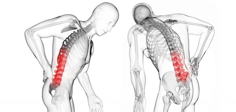
Contraindications for cervical osteochondrosis
- Sleeping on a very hard or soft mattress or high pillow;
- Lift weights; if you need to lift something heavy, you need to straighten your back and bend your knees;
- shoulder bag;
- When the condition worsens, actively move the head and neck;
- Smoking and drinking;
- Do not wear a scarf when walking in cold weather, sit in a ventilated place or near an air conditioner;
- Remaining in uncomfortable positions or sitting for long periods of time;
- wear high heel shoes;
- Break your neck.

prevent disease
- Get rid of excess weight;
- Do gymnastics, swimming, yoga, and dancing every morning;
- Spending time outdoors, especially morning walks, can be helpful;
- Eat right, control salt intake, and follow drinking habits;
- When working sedentary, do a neck warm-up every hour and pay attention to your posture;
- Keep your neck warm;
- Get enough sleep to avoid physical, mental and emotional fatigue.


